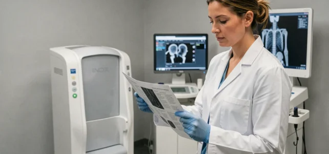
When a doctor prescribes a bone density test, most patients imagine it as a simple examination of the bones. Recent devices installed in medical imaging centers no longer just measure the strength of the skeleton: they also analyze the distribution of muscles and fats in a single acquisition. This evolution is transforming radiology in Aix and the way practitioners monitor their patients.
Next-Generation DEXA: An Examination That Goes Beyond Bone Density
The bone density test relies on DEXA technology (dual-energy X-ray absorptiometry). The principle is simple: two beams of X-rays pass through the body. Depending on the density of the tissues traversed, the device precisely calculates the bone mineral mass, particularly in the lumbar spine and the femoral neck.
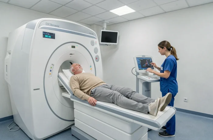
Since the summer of 2026, a new generation of bone densitometers has begun to be deployed in French clinics. These devices perform, in a single acquisition, an assessment of bones, muscles, and fats. In practice, the patient lies down once on the examination table and leaves with three types of data instead of just one.
Why does this evolution matter for the patient? Because a bone weakened by osteoporosis is only part of the problem. The loss of muscle mass (sarcopenia) increases the risk of falls, and thus fractures. Practitioners monitoring bone density and radiology in Aix en Provence now have a tool that intersects these two parameters, refining care well beyond just post-menopausal screening.
Sarcopenia and Body Composition: What the DEXA Exam Reveals to Doctors
Sarcopenia affects an increasing portion of the aging population. Long diagnosed through clinical tests (grip strength, walking speed), it can now be objectively assessed through imaging. The bone densitometer measures appendicular muscle mass, meaning that of the arms and legs, with a precision that biological assessments alone cannot provide.
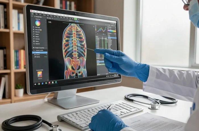
For a general practitioner, this changes the game. Rather than prescribing two separate tests (one for bones, one for body composition), they direct the patient to a single DEXA session. The radiologist produces a report that covers both dimensions.
Who is Concerned by This Expanded Assessment
- Postmenopausal women being monitored for osteoporosis, where muscle loss exacerbates the risk of vertebral or femoral neck fractures
- Patients over 65 with a history of falls, for whom the intersection of bone density and muscle mass guides prevention
- Athletes in longitudinal follow-up, particularly in endurance disciplines where body composition changes rapidly
This positioning of bone densitometry at the crossroads of several specialties (rheumatology, geriatrics, sports medicine) represents a turning point for imaging centers investing in these devices.
Bone Densitometry Market in France: A Re-equipment Underway
DEXA technology is evolving faster than traditional examination sheets suggest. The French manufacturer DMS recorded a significant increase in its bone densitometry activity in the first half of 2026, with revenue growth in this segment compared to 2025 and a strong rebound in the second quarter.
This dynamic reflects a movement of re-equipment in clinics and hospitals. When a center replaces an old bone densitometer with a new model, it gains access to the body composition features mentioned earlier. The installed base is modernizing, and patients benefit directly: faster exams, reduced radiation doses, more detailed reports.
What This Changes for Radiology Centers in Aix
Aix-en-Provence has several medical imaging facilities spread across the Private Hospital of Provence, city radiology offices, and peripheral clinics. Local competition drives each center to differentiate itself.
A next-generation DEXA device becomes both a medical and logistical argument. A multidimensional examination replaces two separate appointments, which reduces the time to care for the patient and frees up slots for the center.
Fracture Prevention in the Elderly: The Role of the General Practitioner
The HAS reminds us that falls in the elderly are not a foregone conclusion. The general practitioner plays a central role in identifying at-risk patients. Bone densitometry is part of this prevention chain, but it is not sufficient on its own.
The DEXA assessment provides a T-score, which compares the patient’s bone density to that of a healthy young adult. A score below -2.5 confirms osteoporosis. However, two patients with the same T-score do not have the same fracture risk if one has good muscle mass and the other does not.
This is precisely where recent devices provide additional information. The T-score alone does not predict the risk of falls: the analysis of body composition completes the clinical picture. The general practitioner can then adjust their recommendations (targeted physical activity, supplementation, referral to a geriatrician).
- Prescription of a bone density test with body composition analysis for identified at-risk patients
- Regular follow-up in the same center, with the same protocol, to compare results over time
- Coordination between radiologist, rheumatologist, and primary care physician for a coherent care pathway
The imaging centers in Aix-en-Provence that integrate these expanded examinations participate in a more comprehensive preventive approach than just osteoporosis screening. The DEXA examination becomes a tool for global prevention, not just a bone diagnosis.